For your convenience, you can easily book your appointment online.
Brooklyn Heights Dental® has helped many in the Brooklyn, NY, community achieve optimal oral and overall health since 1956. Dr. Eugene D. Stanislaus, Dr. Lisa Reid, Dr. Mariam Vonderheide, and Dr. Penny Planzos are highly skilled, compassionate doctors who provide a wide range of dental work, including advanced restorative, general, implant, and cosmetic dentistry procedures. Our team is dedicated to providing the customized care you need at our dental office in Brooklyn Heights.
Dr. Eugene D. Stanislaus is proud to offer the advanced Chao Pinhole Surgical Technique, a suture-free, scalpel-free procedure for gum recession.
To maintain good health, it is important to visit the dentist at least twice a year. At Brooklyn Heights Dental® in Brooklyn, NY, our dentists can provide excellent general, restorative, and cosmetic dentistry in an office that combines a proud legacy with state-of-the-art technology and procedures.
If you haven't seen a dentist in a while, your teeth probably need attention. Regular dental checkups are important because they provide our dentist an opportunity to notice dental or periodontal issues in their earliest stages before they become too serious. They also allow our hygienists to provide a thorough and efficient cleaning in a friendly and calming environment.
We welcome patients from Fort Greene, all of Brooklyn, and throughout greater NYC. We can help you maintain optimal oral health. To learn more or to schedule an appointment at our Brooklyn Heights dental practice, contact us online or call:
Kindarella
Brooklyn, NY
2023
Amazing service and care!! My experience over the last several years has continuously been optimal. Everyone is always so friendly, considerate, professional and thorough.
View on Googleleslie s.
Brooklyn, NY
2023
I am a long-term client of the practice. As always, I received attentive care for my semi-annual cleaning and examination.
View on GoogleWe are committed to providing comprehensive dental care to patients. All our treatments are driven by conservatism, aesthetics, and comfort. Brooklyn Heights Dental®

Dr. Eugene D. Stanislaus is a highly trained second-generation dentist. He has operated Brooklyn Heights Dental® ever since acquiring the practice from his father in 1985. Dr. Stanislaus has studied advanced dental techniques with world-renowned doctors at many prestigious institutes, and he is affiliated with several high-profile professional organizations.

As a prosthodontist, Dr. Lisa Reid specializes in restoring lost or damaged teeth. She is an expert in dental implants and loves to work with patients who want to improve the appearance and function of their smile. In addition to her experience working in private practice, she is also an educator of future dentists, having held positions at the University of Iowa and NYU.
Dr. Vonderheide received her doctor of dental medicine (DMD) degree with clinical honors from the University of Pennsylvania School of Dental Medicine. Upon graduation from Penn, Dr. Vonderheide completed a three-year residency in postgraduate prosthodontics at Montefiore Medical Center in the Bronx.
Dr. Penny Planzos attended the University of Pennsylvania School of Dental Medicine and graduated with clinical honors. She went on to pursue advanced training in Prosthodontics at Columbia University College of Dental Medicine, where she focused on full mouth rehabilitation, complex dental treatment, and cosmetics.
The tradition at Brooklyn Heights Dental® dates back more than five decades. Dr. Stanislaus' father, Dr. Lamuel Stanislaus, initially opened the practice in 1956, just three miles from our current location. Our commitment today is the same as it was then. We are focused on delivering personalized attention and exceptional care to help members of the community achieve the very best smile possible. We are committed to treating our patients just as we would treat a member of our own family.
We welcome patients from Brooklyn Heights, Fort Greene, all of Brooklyn, and throughout greater NYC. Whether you are looking to address cosmetic issues with your smile, need attention for a toothache or gum infection, or just are looking for a new dentist, we are happy to welcome you to our friendly office. Come experience what sets our family dental practice apart. To schedule an appointment at our dental practice in Brooklyn Heights, contact us online or call:
Myrtle Kennedy
Brooklyn, NY
2023
I love my dental hygienist and my dentist. They are so helpful and knowledgeable. Thanks for caring.
View on GoogleCorey Shaun Run
Brooklyn, NY
2023
Friendly and professional staff that are always on their A game. It's better than any other dental office I've been to.
View on GoogleIf you have missing teeth and want to reclaim the functionality and other benefits of your natural teeth, we offer dental implants. These titanium posts are placed in the jaw, providing a stable foundation for your crowns, bridges, or dentures. With the right care, implants can last for decades. By performing every stage of the procedure at our dental practice, from surgical placement to the attachment of the final restoration, we can provide reliable and convenient care from start to finish.
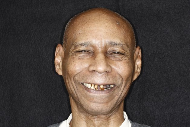
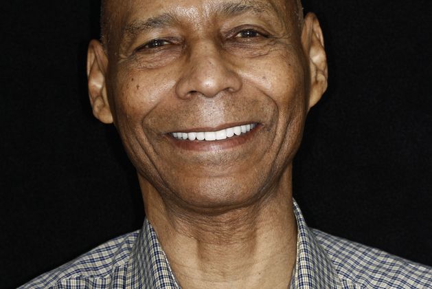
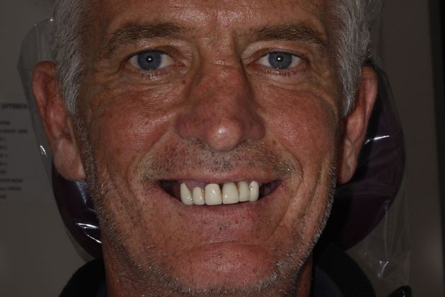
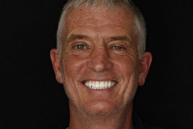
Teeth that are chipped, cracked, discolored, or otherwise imperfect can take a serious toll on your confidence. A porcelain veneer is a great solution if you want to cover a less-than-perfect tooth. A veneer is a thin shell that your dentist can bond to the front surface of a tooth to create a look of symmetry and health. At Brooklyn Heights Dental® in Brooklyn, NY, our veneers are custom-made of medical-grade ceramic that is carefully crafted to match the appearance of natural teeth.
If you would like a straighter smile but are put off by the thought of metal braces, Invisalign® is a discreet alternative orthodontic treatment. The aligners are practically invisible against your smile, and each one is made of a smooth plastic that does not irritate your cheeks and gums. Additionally, the removable aligners offer no restrictions on diet or dental care. You can continue to enjoy your favorite foods while maintaining optimal oral health.
A painful tooth that has been weakened by infection can be saved by root canal therapy. Virtually painless, a root canal removes the infected pulp of the tooth and preserves the exterior structure. Furthermore, root canal therapy can eliminate the need for extraction by reinforcing the tooth with a dental crown. If you suspect you have an infected tooth, we advise you to contact us as soon as possible to avoid the development and spread of serious health issues.
A dental crown acts like a cap and covers a damaged tooth. The dentists at our Brooklyn, NY, dental practice pride themselves on crafting crowns that are aesthetically pleasing while also being strong and durable. Because of our CEREC technology, many patients can take advantage of same-day crowns that are crafted and placed in just one visit.
We provide a full range of procedures to meet your varied needs and help you achieve a smile you can feel proud to share.
Thanks to advanced dental technology, there's no reason to settle for a smile you don't love. Our doctors have decades of experience in cosmetic and restorative dentistry, and they can help you achieve the beautiful smile you want, whether your teeth are crooked, stained, broken, or missing entirely.
Our gentle and caring team will ensure your appointment is comfortable. If the thought of visiting the dentist makes you nervous, we can put your worries at ease with sedation dentistry. We make every effort to ensure you have a pleasant time at our dental practice.
To learn more about how restorative and cosmetic dentistry can transform your smile, request a consultation at our Brooklyn Heights office by contacting us online or calling:
Denise C.
Brooklyn, NY
2023
There all so welcoming. I went in for the zoom whitening. I was a little scared but honestly they informed me about everything. As soon as I saw my teeth i fell in love . They came out beautiful. Shout out to Andria you are the best. You made me very comfortable, educated me about the whole process. Thank you for being patient with me . Awesome place !!! I highly recommend . I’ll definitely be back .
View on GoogleShanoya Belcher
Brooklyn, NY
2022
I don't normally post reviews, but this experience has been one of the best ones yet. From the moment, I called and I spoke to Theresa it was just amazing. I walked in and was greeted so warmly by the staff. I received the best cleaning from Jonathan who explained EVERYTHING to me. They were so warm and friendly. The Dentist was the nicest man ever. His bed side manners was top tier. I have finally found a Dentist office that treated me like I belonged there. Not to mention I received a little gift bag too. Thank you guys!
View on GoogleEven if you have the best oral hygiene, eating and drinking can lead to discolored and stained teeth. Professional teeth whitening can quickly remove the toughest stains. In a single appointment, you can experience a transformation that can't be matched by over-the-counter alternatives.
To help you achieve a brighter smile, we use the latest innovative technology. Our dentists provide Zoom!® teeth whitening treatments at our dental office in Brooklyn Heights. Providing stunning results, Zoom! can give you a whiter smile in just one 45-minute visit. Contact our dental practice today for more information about how we can boost your confidence with professional teeth whitening treatment.

Gingivitis, the earliest stage of gum disease, is characterized by swollen and red gums that may bleed, especially when flossing. If left untreated, gingivitis can develop into periodontitis, which can lead to tooth loss. If this wasn't bad enough, recent research shows a direct link between periodontal disease and cardiovascular disease. Researchers think that the bacteria in gum disease can travel throughout the body, triggering inflammation in the heart’s vessels and infection in heart valves. Fortunately, our dentists offer effective, state-of-the-art laser dentistry for gum disease.
Dr. Stanislaus is one of very few dentists in New York City certified to perform the Laser Assisted New Attachment Procedure (LANAP), which has benefited many of our neighbors in Brooklyn Heights. LANAP is a technologically advanced treatment for gum disease that involves the use of a laser to remove diseased gum tissue. Dr. Stanislaus believes that LANAP is the best available procedure to address periodontal disease.

Periodontal disease is unfortunately common. In fact, the CDC has estimated that half of Americans 30 and older are suffering from some form of gum disease.
Nia
Brooklyn, NY
2022
Professional. Patient. Knowledgeable. Informative. These are just a few of the words I could use to describe the Brooklyn Heights Dental staff. My appointments have been wonderful and I’m not a person who enjoys the dentist for the same reasons most people don’t but I can say without a shadow of doubt that this is the best practice I’ve been to EVER and they are so welcoming and skilled. It baffles me how I literally picked them on a random day off google!! Thank you so much!!
View on GoogleUlysses Williams
Brooklyn, NY
2022
The staff was friendly and made it easy to make the initial appointment. In addition they took the time to explain the projected costs and such for the subsequent upcoming visits. The hygienist and dentist were friendly, knowledgeable, and consistently exhibited the highest degree of professionalism. Moreover, they explained the complicated matters to me in a manner that I could understand. I look forward to seeing this Team again for my upcoming visits.
View on GoogleThe team at Brooklyn Heights Dental® has been committed to caring for our community's smiles since 1956. Dr. Eugene D. Stanislaus and Dr. Lisa Reid are affiliated with several renowned institutions and organizations, including:
To set up an appointment or learn more about our services, give us a call at (718) 857-6639 or contact us online.
"I've been very happy with my experiences at Dr. Stanislaus's office, and highly recommend him to anyone in the New York City area. He and his staff go above and beyond to do the best job possible." -R.B. (Patient)
©2008 - 2024 Brooklyn Heights Dental, P.C. | Forever Website® 2.0 | Designed & Developed by Einstein Dental